Exploring Anatomy & Physiology in the Laboratory 4th Edition: A Comprehensive Plan
This edition emphasizes interdisciplinary knowledge for future physicians‚ requiring educators to foster cognitive integration of information throughout medical school curricula․
This fourth edition of “Exploring Anatomy & Physiology in the Laboratory” builds upon established foundations‚ recognizing the crucial need for physicians to synthesize knowledge across various disciplines․ The modern medical landscape demands more than rote memorization; it necessitates a deep‚ interconnected understanding of biological systems․
Consequently‚ this edition prioritizes opportunities for students to actively integrate information‚ mirroring the cognitive demands of clinical practice․ Educators are challenged to move beyond traditional siloed learning and cultivate a holistic approach to medical education․ This edition aims to equip students with the skills to effectively apply anatomical and physiological principles in real-world scenarios‚ fostering critical thinking and problem-solving abilities․
II․ Core Principles of Anatomy & Physiology
Fundamental to medical training is the integration of anatomy and physiology – structure dictates function‚ and function relies on structure․ This edition reinforces this core principle‚ emphasizing the interconnectedness of bodily systems․ Effective physicians require a robust understanding of how these systems collaborate to maintain homeostasis․
The text promotes cognitive integration‚ moving beyond isolated facts to demonstrate how anatomical features enable specific physiological processes․ This approach prepares students for the complex‚ interdisciplinary challenges encountered in clinical settings‚ demanding a holistic grasp of biological mechanisms and their clinical relevance․
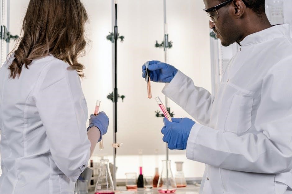
III․ Laboratory Safety Protocols
Prioritizing a secure learning environment is paramount in anatomy and physiology labs․ This edition underscores the necessity of comprehensive safety training for all students‚ aligning with the demands of modern medical education․ Educators must instill a culture of responsibility and awareness regarding potential hazards․
Effective training facilitates cognitive integration of safety procedures alongside scientific exploration․ Protocols encompass handling biological specimens‚ proper use of dissection tools‚ and adherence to general lab rules‚ ensuring both student and instructor well-being throughout the practical learning experience․
III․A․ General Safety Rules
Establishing foundational safety habits is crucial for a productive lab experience․ Students must consistently wear appropriate personal protective equipment – lab coats‚ gloves‚ and eye protection – to minimize exposure risks․ Maintaining a clean and organized workspace prevents accidents and contamination․
Food and drink are strictly prohibited within the lab to avoid accidental ingestion of potentially hazardous materials․ Proper handwashing protocols before and after handling specimens are essential․ Adherence to these rules fosters a responsible learning environment‚ supporting the integration of safe practices alongside anatomical study․
III․B․ Handling Biological Specimens
Respectful and cautious handling of biological specimens is paramount; All materials should be treated as potentially infectious‚ regardless of preservation methods․ Proper disposal procedures‚ following established biohazard protocols‚ are non-negotiable․ Specimens must be handled with designated tools – forceps‚ probes – avoiding direct contact with skin․
Detailed labeling and accurate record-keeping are vital for traceability and preventing mix-ups․ Students should be thoroughly trained on specimen preservation techniques and understand the risks associated with each material‚ ensuring responsible integration with anatomical studies․
III․C․ Use of Dissection Tools
Proficient and safe utilization of dissection tools is crucial for effective anatomical study․ Students require comprehensive training on proper handling of scalpels‚ scissors‚ probes‚ and forceps‚ emphasizing controlled movements and awareness of surrounding tissues․ Sharp instruments demand respect; always cut away from the body․
Regular inspection of tools for damage is essential‚ and reporting any defects is mandatory․ Proper cleaning and storage protocols must be strictly followed to maintain tool integrity and prevent accidents‚ fostering responsible integration within laboratory practices․
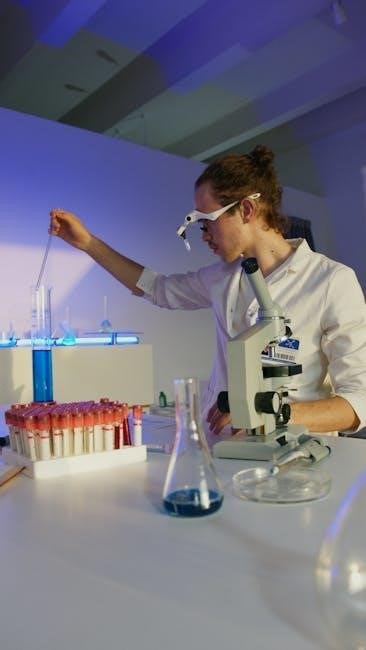
IV․ Histology & Microscopy
Histological study provides a microscopic view of tissues‚ fundamental to understanding organ structure and function․ This section details tissue preparation – fixation‚ embedding‚ sectioning‚ and staining – vital for clear visualization․ Common staining techniques like Hematoxylin and Eosin (H&E) and Trichrome reveal cellular details․
Students will learn to identify the four primary tissue types: epithelial‚ connective‚ muscle‚ and nervous‚ under the microscope․ Accurate observation and interpretation are key‚ building a strong foundation for integrated physiological understanding․
IV․A․ Tissue Preparation Techniques
Proper tissue preparation is crucial for high-quality microscopic examination․ This involves several key steps: fixation to preserve structure‚ embedding in paraffin for support‚ and microtomy to create thin sections․ These sections are then mounted on slides for staining and observation․
Detailed protocols for each technique will be provided‚ emphasizing the importance of precise timing and reagent use․ Students will practice these methods‚ learning to troubleshoot common issues and ensure optimal tissue preservation for accurate histological analysis․
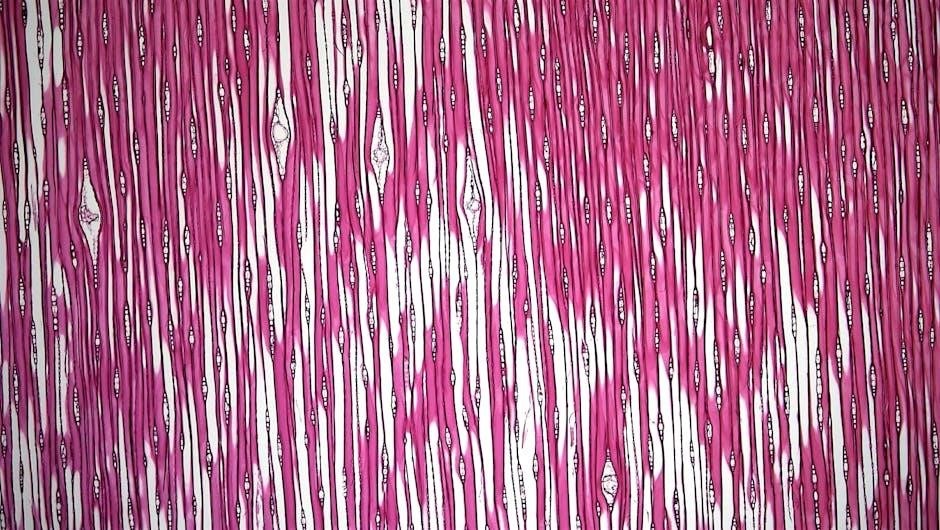
IV․B․ Staining Methods (H&E‚ Trichrome)
Histological staining enhances contrast‚ revealing cellular details․ Hematoxylin and Eosin (H&E) is the most common stain‚ differentiating nuclei (blue) from cytoplasm and extracellular matrix (pink)․ Masson’s Trichrome stains collagen blue‚ aiding in identifying fibrosis․
This lab will cover the principles behind each stain‚ including mordants and dye chemistry․ Students will practice staining techniques‚ learning to optimize color development and interpret staining patterns to identify different tissue components accurately․ Proper handling of staining reagents is paramount․
IV․C; Identifying Tissue Types Under the Microscope
Microscopic examination is crucial for tissue identification․ Students will learn to differentiate the four primary tissue types: epithelial‚ connective‚ muscle‚ and nervous․ Recognizing cellular arrangements‚ matrix composition‚ and specialized structures is key․
This section focuses on correlating histological features with tissue function․ Practical exercises involve identifying tissues from prepared slides‚ utilizing staining characteristics observed with H&E and Trichrome․ Emphasis will be placed on accurate terminology and descriptive analysis‚ building a strong foundation for organ system study․
V․ Anatomical Dissection – General Overview
Dissection provides a hands-on understanding of anatomical relationships․ This section outlines the principles of respectful and methodical dissection‚ emphasizing careful tissue separation and identification of structures․ Students will learn proper techniques for preserving specimens and documenting findings․
A strong emphasis is placed on correlating anatomical observations with physiological function‚ reinforcing the integrated nature of anatomy and physiology․ This overview prepares students for systematic dissection of various organ systems‚ fostering spatial reasoning and anatomical vocabulary․
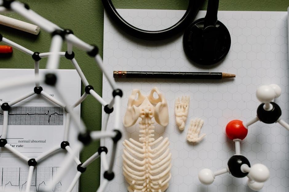
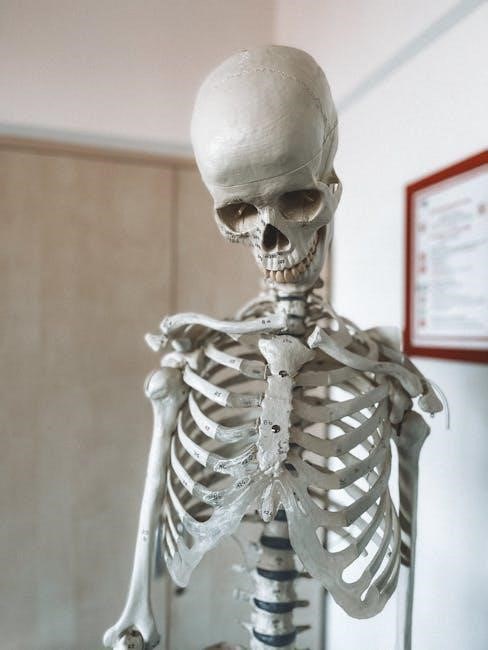
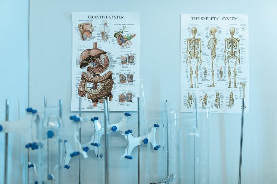
VI․ Skeletal System Dissection
This module focuses on the detailed dissection of the skeletal system‚ beginning with bone identification and landmark recognition․ Students will explore different bone classifications and articulate the functions of skeletal markings․ Practical exercises involve identifying various joint types – fibrous‚ cartilaginous‚ and synovial – and analyzing their articulation․
Emphasis is placed on understanding the relationship between bone structure and biomechanical function․ Students will learn to assess skeletal variations and apply this knowledge to clinical scenarios‚ solidifying their grasp of anatomical principles․
VI․A․ Bone Identification & Markings
This section centers on mastering bone identification through hands-on dissection and skeletal models․ Students will systematically identify individual bones‚ categorizing them by region (axial vs․ appendicular) and shape․ A core component involves recognizing key bone markings – processes‚ foramina‚ fossae‚ and tubercles – and correlating these features with their specific functions․
Understanding the purpose of these markings is crucial for comprehending muscle attachment sites‚ nerve pathways‚ and blood vessel routes․ Practical exercises reinforce this knowledge‚ preparing students for advanced anatomical studies․
VI․B․ Joint Types and Articulation
This module delves into the classification of joints based on their structural and functional characteristics․ Students will differentiate between fibrous‚ cartilaginous‚ and synovial joints‚ analyzing their unique components and range of motion․ Dissection exercises will focus on identifying ligaments‚ menisci‚ and articular cartilage‚ understanding their roles in joint stability and movement․
Emphasis will be placed on correlating joint structure with specific movements‚ preparing students to analyze biomechanical principles and clinical implications of joint dysfunction․
VII․ Muscular System Dissection
This section provides a hands-on exploration of skeletal muscle anatomy‚ focusing on identification of major muscles‚ their origins‚ insertions‚ and actions․ Dissection will reveal muscle fiber arrangement and fascial coverings‚ illustrating how these structures contribute to force generation and movement․ Students will learn to differentiate between agonist‚ antagonist‚ and synergist muscle groups․
The lab will reinforce understanding of how integrated knowledge across disciplines is crucial for physicians‚ linking muscle function to overall physiological processes․
VII․A․ Muscle Identification & Function
This lab centers on accurately identifying superficial and deep muscles of the body‚ utilizing anatomical landmarks and dissection techniques․ Students will correlate muscle names with their specific actions – flexion‚ extension‚ abduction‚ adduction‚ and rotation – observing how these movements contribute to complex physiological functions․
Emphasis is placed on understanding how physicians require integrated knowledge; therefore‚ students will explore the clinical relevance of muscle injuries and disorders‚ linking anatomical structure to functional impairment․
VII․B․ Muscle Fiber Types & Characteristics
This section delves into the microscopic anatomy of skeletal muscle‚ differentiating between Type I (slow-twitch)‚ Type IIa‚ and Type IIx (fast-twitch) muscle fibers․ Students will analyze histological slides‚ identifying key characteristics like fiber diameter‚ myoglobin content‚ and capillary density‚ relating these features to metabolic properties and functional capabilities․
Understanding these distinctions is crucial‚ as physicians must integrate knowledge across disciplines; therefore‚ the lab connects fiber type composition to athletic performance and disease states․
VIII․ Nervous System Exploration
This module focuses on the intricate structures of the nervous system‚ beginning with a detailed brain dissection․ Students will identify major brain regions – cerebrum‚ cerebellum‚ brainstem – and associated structures‚ correlating anatomical features with neurological functions․
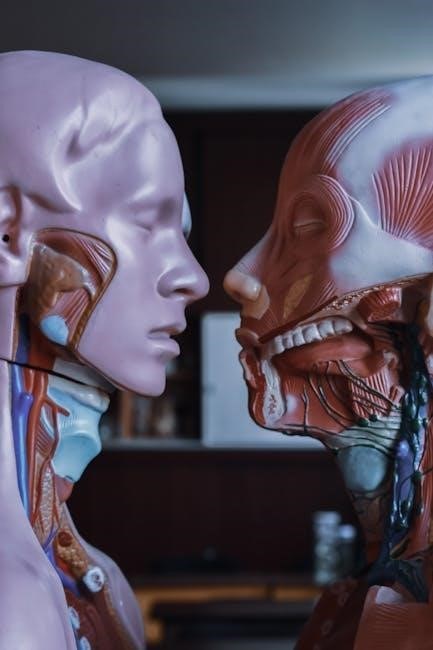
The lab emphasizes the need for physicians to integrate knowledge‚ requiring students to connect anatomical observations to clinical scenarios․ Furthermore‚ spinal cord anatomy and nerve pathways will be explored‚ reinforcing the importance of interdisciplinary understanding within medical education․
VIII․A․ Brain Dissection & Structures
This lab provides a hands-on experience with sheep brain dissection‚ allowing students to identify key anatomical landmarks․ Emphasis is placed on correlating structure with function‚ understanding how each region contributes to neurological processes․ Students will meticulously dissect and observe the cerebrum‚ cerebellum‚ and brainstem‚ noting gyri‚ sulci‚ and major fissures․
The exercise reinforces the need for physicians to integrate knowledge‚ connecting anatomical observations to clinical applications․ This detailed exploration fosters a deeper understanding of the nervous system’s complexity and its vital role in overall health․
VIII․B․ Spinal Cord Anatomy & Nerve Pathways
The laboratory focuses on dissecting and identifying the external and internal structures of the spinal cord․ Students will trace the pathways of ascending and descending nerve tracts‚ understanding their roles in sensory and motor functions․ Observing the gray and white matter organization is crucial‚ correlating it with reflex arcs and neurological control․
This practical experience reinforces the importance of integrated knowledge‚ preparing future physicians to diagnose and treat neurological conditions effectively․ Understanding nerve pathways is fundamental to clinical practice․
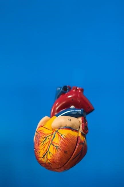
IX․ Cardiovascular System Study
This section emphasizes the interconnectedness of anatomical structures and physiological functions within the circulatory system․ Students will perform heart dissections‚ meticulously tracing blood flow through chambers‚ valves‚ and major vessels․ Identifying coronary arteries and understanding their clinical significance is paramount․
Furthermore‚ the lab explores blood vessel structure‚ relating it to hemodynamics․ This hands-on approach reinforces the need for physicians to integrate knowledge across disciplines for effective patient care and diagnosis․
IX․A․ Heart Dissection & Blood Flow
Detailed heart dissections are central‚ allowing students to physically trace blood’s pathway․ Identifying chambers‚ valves (tricuspid‚ mitral‚ pulmonary‚ aortic)‚ and major vessels (vena cava‚ aorta‚ pulmonary artery/vein) is crucial․ Emphasis is placed on correlating anatomical structures with their specific physiological roles in circulation․
Understanding coronary artery location and potential blockage scenarios prepares students for clinical applications․ This reinforces the need for integrated knowledge‚ vital for future physicians’ diagnostic and treatment capabilities․
IX․B․ Blood Vessel Identification & Structure
Comprehensive study of arterial‚ venous‚ and capillary structures is paramount․ Students will differentiate between vessel types based on wall thickness‚ lumen size‚ and valve presence․ Identifying major arteries (aorta‚ femoral‚ carotid) and veins (vena cava‚ jugular) is essential․
The lab emphasizes how structural differences relate to blood pressure and flow dynamics․ This reinforces the importance of integrated knowledge‚ preparing future physicians to understand cardiovascular pathologies and treatment strategies effectively․
X․ Respiratory System Investigation
This section focuses on the anatomical structures facilitating gas exchange․ Students will dissect lungs‚ tracing the pathway of air from the nasal cavity to the alveoli․ Examining the diaphragm and intercostal muscles demonstrates the mechanics of breathing․
Labs incorporate spirometry to measure lung volumes and capacities‚ linking structure to function․ Understanding these principles is crucial‚ as physicians require integrated knowledge to diagnose and manage respiratory conditions effectively‚ bridging anatomical understanding with physiological processes․
XI․ Digestive System Examination
This module explores the anatomy of the alimentary canal‚ from the mouth to the anus‚ dissecting organs like the stomach‚ small intestine‚ and liver․ Students will identify key structures involved in mechanical and chemical digestion‚ observing peristalsis and enzymatic activity․
Labs emphasize the integrated nature of physiological processes‚ mirroring the demands placed on physicians․ Understanding how anatomical structures support digestion is vital for diagnosing and treating related disorders‚ requiring a comprehensive‚ interdisciplinary approach to medical education․
XII․ Urinary System Analysis
This section focuses on the kidneys‚ ureters‚ bladder‚ and urethra‚ dissecting to reveal nephron structure and function․ Students will analyze urine samples‚ identifying key components and relating them to physiological processes like filtration‚ reabsorption‚ and secretion․
Labs reinforce the need for physicians to integrate knowledge across disciplines‚ understanding how the urinary system impacts overall homeostasis․ This integrated approach is crucial for diagnosing and managing conditions affecting fluid balance and waste removal‚ demanding a holistic educational perspective․
XIII․ Reproductive System Study
This module delves into the male and female reproductive anatomies‚ emphasizing gametogenesis and hormonal control․ Dissections will highlight structures crucial for reproduction‚ while laboratory exercises explore the physiological basis of the menstrual cycle and spermatogenesis;
The text stresses the importance of integrated knowledge for physicians‚ requiring students to connect reproductive physiology with broader systemic functions․ Educators must provide opportunities for cognitive integration‚ preparing students for clinical scenarios involving reproductive health and endocrine imbalances․
XIV․ Sensory Organ Dissection
This section focuses on the anatomy of the eye‚ ear‚ nose‚ and tongue‚ examining structures responsible for sight‚ hearing‚ smell‚ and taste․ Dissections will reveal the intricate pathways of sensory information‚ while microscopy explores the cellular components of sensory receptors․
The 4th edition underscores the need for physicians to integrate knowledge across disciplines․ Therefore‚ educators should facilitate cognitive connections between sensory organ anatomy‚ physiology‚ and neurological pathways‚ preparing students for clinical applications involving sensory deficits and neurological assessments․
XV․ Utilizing Virtual Labs & Digital Resources
This module introduces students to cutting-edge virtual dissection tools and interactive 3D anatomical models‚ supplementing traditional lab experiences․ Digital resources‚ including online quizzes and interactive tutorials‚ reinforce key concepts and provide personalized learning opportunities․
The 4th edition recognizes the importance of interdisciplinary knowledge․ Educators should leverage these digital tools to encourage students to cognitively integrate anatomical structures with physiological functions‚ mirroring the complex problem-solving required in clinical practice and enhancing overall comprehension․
XVI․ Physiological Measurements & Data Analysis
This section focuses on developing students’ abilities to collect‚ record‚ and analyze physiological data obtained from laboratory experiments․ Emphasis is placed on understanding the correlation between anatomical structures and their functional outputs‚ utilizing techniques like heart rate monitoring and respiratory volume measurements․
The 4th edition stresses the need for physicians to integrate knowledge․ Students will learn to interpret data‚ draw conclusions‚ and apply these findings to understand complex physiological processes‚ mirroring the analytical skills crucial for effective clinical decision-making․
XVII․ Common Laboratory Exercises & Experiments
This component details a range of practical exercises designed to reinforce anatomical and physiological concepts․ These include dissections‚ microscopy‚ and physiological measurements‚ all geared towards building a strong foundation for future medical professionals․
The 4th edition acknowledges the importance of integrated learning‚ mirroring the demands placed on physicians․ Experiments will encourage students to connect theoretical knowledge with hands-on experience‚ fostering a deeper understanding of how anatomical structures contribute to overall physiological function and clinical scenarios․
XVIII․ Troubleshooting Common Dissection Challenges
This section addresses frequent difficulties encountered during dissection‚ offering practical solutions and guidance for students․ Recognizing that effective learning requires overcoming obstacles‚ the 4th edition provides detailed advice on handling delicate tissues‚ identifying key structures‚ and managing unexpected anatomical variations․
It emphasizes the need for educators to prepare students for the complexities of real-world anatomy‚ aligning with the integrated knowledge base physicians require․ Strategies for collaborative problem-solving and resource utilization are also highlighted‚ promoting independent learning․
XIX․ Assessment & Grading in the Anatomy & Physiology Lab
Effective assessment is crucial for gauging student comprehension and skill development․ This edition advocates for diverse evaluation methods beyond traditional quizzes‚ including practical exams‚ dissection accuracy assessments‚ and collaborative project work․ Grading rubrics should clearly define expectations‚ aligning with the integrated knowledge physicians need․
The text stresses that educators must provide constructive feedback‚ fostering a learning environment where students can refine their anatomical understanding and prepare for clinical applications․ Emphasis is placed on evaluating cognitive integration․
XX․ Integration with Clinical Applications
This edition strongly advocates bridging the gap between foundational anatomical knowledge and real-world clinical scenarios․ Students must learn to apply their understanding of anatomy and physiology to diagnose and treat medical conditions․ Case studies‚ simulated patient encounters‚ and discussions of clinical correlations are vital components․
The text highlights the necessity for physicians to integrate knowledge across disciplines‚ demanding educators provide opportunities for students to cognitively connect lab findings with clinical practice‚ ultimately enhancing patient care․
XXI․ Future Trends in Anatomy & Physiology Education
The evolving landscape of medical education necessitates a continued focus on interdisciplinary learning and cognitive integration․ Future trends will likely emphasize advanced virtual reality simulations‚ augmented reality applications‚ and personalized learning pathways․
Educators must prepare students to navigate complex medical challenges by fostering their ability to synthesize information across disciplines․ This edition underscores the importance of equipping future physicians with the skills needed for lifelong learning and adaptation in a rapidly changing healthcare environment․
Ultimately‚ the successful integration of anatomy and physiology relies on providing students with opportunities to connect theoretical knowledge to practical application․ This edition champions a holistic approach‚ recognizing the physician’s need to synthesize information across multiple disciplines․
By prioritizing cognitive integration and embracing innovative educational tools‚ we empower future healthcare professionals to excel․ Continued emphasis on these principles will ensure a robust and adaptable medical workforce‚ prepared to meet the evolving demands of patient care and medical advancement․